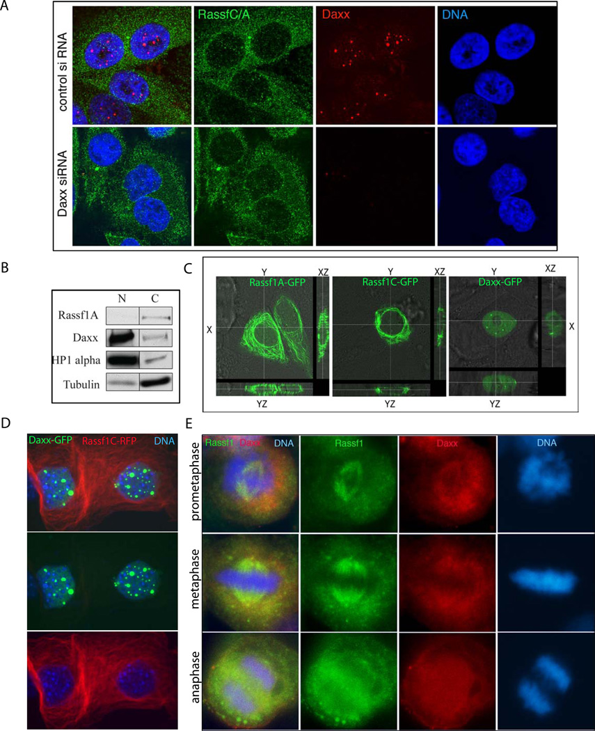
Figure 2. Cell cycle dependent localization of Rassf1 C/A and Daxx.
A) Rassf1C/A and Daxx localization during interphase. Control- (top) and Daxx-depleted (bottom) HEp2 cells were immunostained for Rassf1C/A (green) and Daxx (red); DNA (blue) for nuclear visualization. Note nuclear localization of Daxx in PML NBs (top). Cytoplasmic localization of Rassf1C/A is unaltered upon Daxx depletion (bottom). B) Biochemical fractionation of cells shows differential localization of Daxx/Rassf1 proteins. HEp2 cells were separated into nuclear (N) and cytosolic (C) fractions. Daxx is found in HP1-alpha containing nuclear fractions, while Rassf1 is found in tubulin-containing cytoplasmic fractions. C) Distribution of GFP-Rassf1A, GFP-Rassf1C and GFP-Daxx in HEp2 cells. GFP-Rassf1A (left), -Rassf1C (middle) and -Daxx (right) were transiently transfected into HEp2 cells and then analyzed by confocal microscopy. Note that distribution of both Rassf1 isoforms (Rassf1A and Rassf1C) is exclusively cytoplasmic (compare XY, XZ and YZ planes), while, in contrast, the distribution of Daxx is exclusively nuclear. D) Distribution of Daxx-GFP and Rassf1C-RFP upon double transfection in HEp2 cells. Daxx-GFP is nuclear mostly accumulating in domains (PML NBs), while Rassf1C is cytoplasmic. E) Partial co-localization of Daxx and Rassf1 during mitotic stages. HEp2 cells were immunostained with Rassf1C/A ab (green) and Daxx (red).

