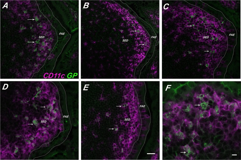
Figure 3. GPs persist within SED DCs.
FITC-GPs were administered to BALB/c mice by gavage, as described in Materials and Methods. PPs were collected at indicated time points thereafter and then cryosectioned, immunostained with anti-CD11c antibodies and viewed by confocal laser scanning microscopy. FITC-GPs are shown in green (arrows) and CD11c+ DCs in magenta (arrow heads). The panels correspond to the following time points (in hours) (A) 2.5; (B) 6; (C) 24; (D) 48; (E) 72 hrs. (F) Close up of SED CD11c+ DCs from tissues taken at 24 hr. In Panels A-E the scale bar corresponds to 50 μm. In Panel F the scale bar is 20 μm.

