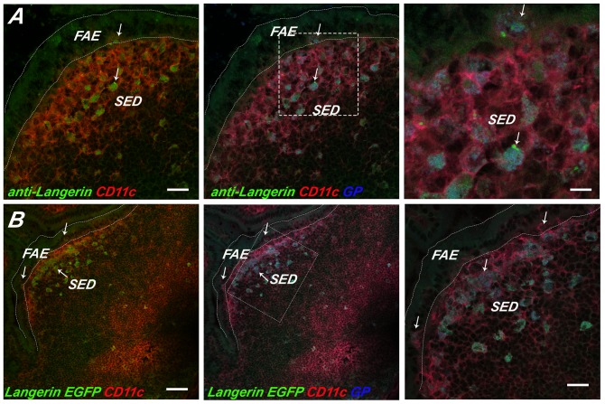
Figure 6. GPs localize within Langerin+ SED DCs.
APC-labeled GPs (blue) were administered to (Panel A) BALB/c or (Panel B) Langerin E-GFP-DTR transgenic mice by gavage, as described in Materials and Methods. PP were collected 24 h later, cryosectioned, immunostained and viewed by three color, confocal laser scanning microscopy. (Panel A) In panel A, BALB/c PP cryosections were immunostained with anti-Langerin mAb RMUL.2 (green; arrows) and anti-CD11c (red). In Panel B, PP cryosections from Langerin-EGFP-DTR transgenic mice were immunostained with anti-CD11c (red) only. In both panels, the left box shows Langerin (green) and CD11c (red) signals only, the middle box shows Langerin (green), CD11c (red), and the APC-GPs (blue) signals, while the right boxes represent a magnification of the dashed square in the middle box. Scale bars correspond to 100 μm (right and middle) or 50 μm.

