
Figure 1.
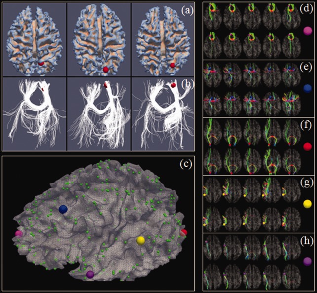
(a) Left precuneus (by the red sphere) in three subjects. (b) Fiber connection profiles of the precuneus in three subjects. (c) The 358 DICCCOL landmarks [Zhu et al., in press]. (d–h) Similar fibers connected to the five landmarks, represented by enlarged bubbles in (c), in two separate groups of five subjects (in two rows), respectively. [Color figure can be viewed in the online issue, which is available at http://wileyonlinelibrary.com.]