
Figure 3.
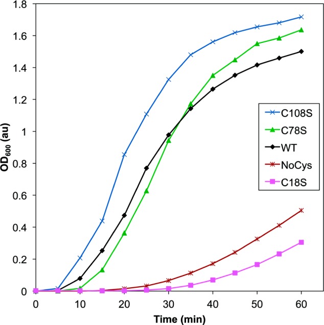
Comparison of UVR-induced aggregation of HγD-Crys constructs with select cysteine residues replaced with serines. Samples contained protein at 1 mg/mL in reaction buffer, and light scattering was monitored at 600 nm as a function of UVR exposure time: WT (black diamonds), C18S (magenta squares), C78S (green triangles), C108S (blue X’s), and NoCys (maroon hashes).