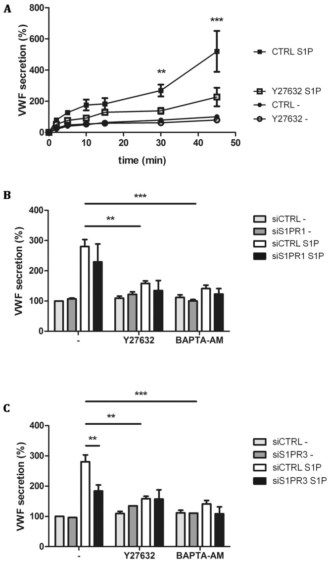
Figure 4. Pharmacological inhibition of S1P-induced VWF secretion.
CTRL and Y27632 treated HUVECs were incubated for indicated times with 1 μM S1P or SF medium alone (-) (A). (B) siCTRL and siS1PR1 treated HUVECs were incubated for 45 minutes with 1 μM S1P or SF medium alone (-) in the presence of 10 μM Rho kinase inhibitor Y27632 or 100 μM calcium chelator BAPTA-AM. (C) siCTRL and siS1PR3 treated HUVECs were incubated for 45 minutes with 1 μM S1P or SF medium alone (-) in the presence of Rho kinase inhibitor Y27632 or calcium inhibitor BAPTA-AM. The amount of VWF secreted in the medium was measured by ELISA; VWF secreted by unstimulated siCTRL treated cells after 45 minutes was set to 100%. Three independent experiments were performed. Statistical significance was assessed by 2-way ANOVA followed by Bonferroni post-hoc test for selected comparison (***P<0.001; **P<0.01 and *P<0.05).

