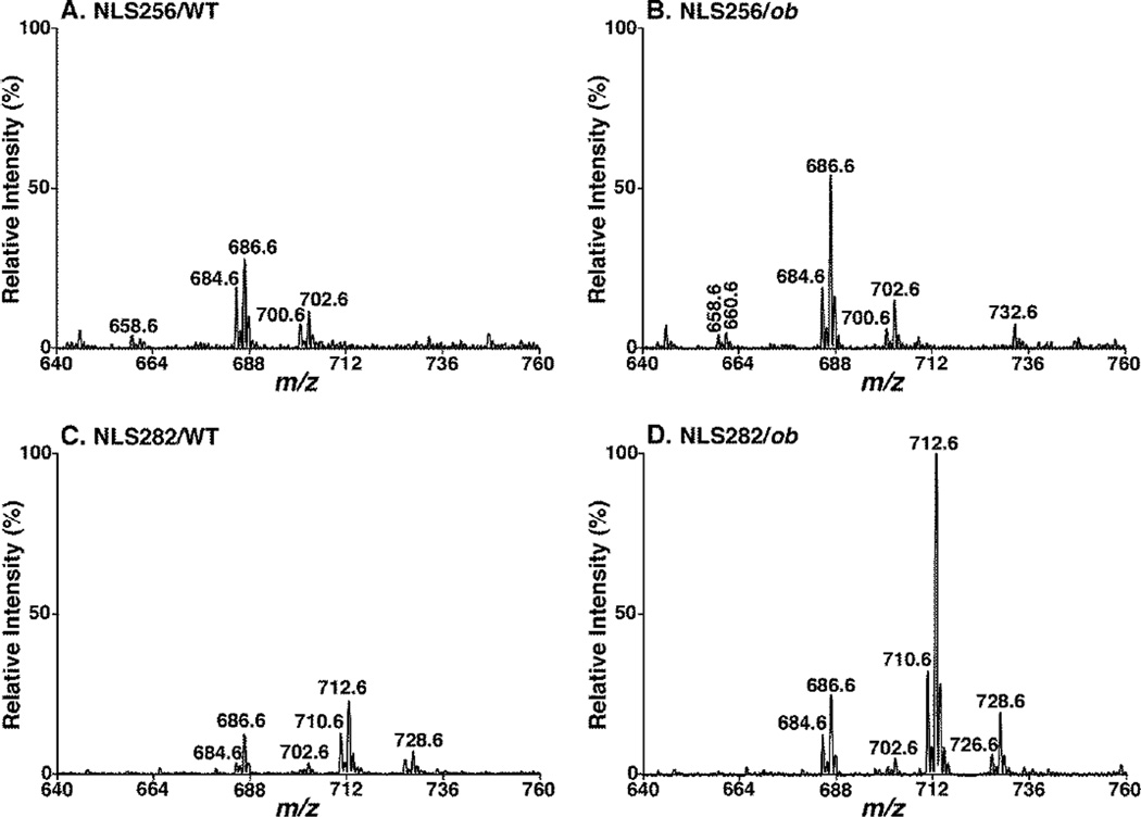
Figure 4.
Comparison of neutral loss scans of fatty acyl chains from lipid extracts of wild type vs. ob/ob liver tissues. Representative MS/MS spectra of NLS256 (i.e., 16:0 FA, A and B) and NLS282 (i.e., 18:1 FA, C and D) were acquired from lipid extracts of WT (A and C) and ob/ob (B and D) mouse liver samples with addition of LiOH at collision energy of 35 eV as described under “MATERIALS AND METHODS”. All the spectra were displayed after separate normalization to the internal standard of each sample and then to the base peak in Panel D. The NLS spectra indicate significantly higher content of DAG species (particularly those containing 18:1 FA) in ob/ob (B and D) compared to WT mouse liver (A and C) at 3 months of age.

