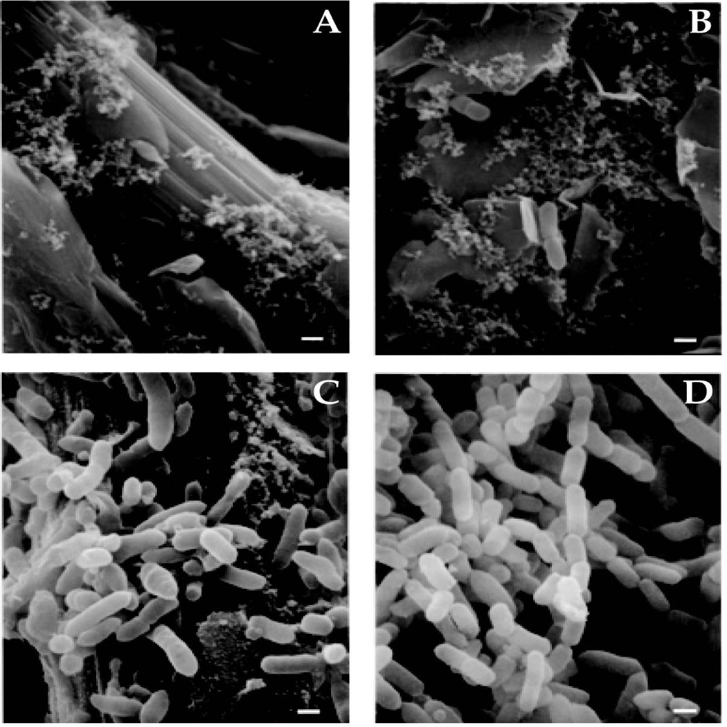
FIGURE 1. Attachment of B. thetaiotaomicron to carbon paper surfaces in the chemostat (A-D).
Scanning electron micrographs of the growth surface sampled at 8 h (panels A and B) and 8 days (panels C and D) after inoculation. Few bacteria are shown in panels A and B, illustrating the rarity of their association with the growth surface at this early time point. Bars, 0.5 µm.

