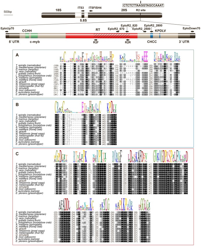
Figure 1. Diagram of the EploR2 insertion site, its molecular structure and alignment with other R2 elements.
The rDNA transcription unit is represented above, with the R2 site indicated. The sequence of the insertion target (box) with a vertical arrow is indicating the exact site of insertion. In the diagram of the R2 element the striped area indicates EploR2_clon3, which was the first sequence obtained with the R2-F and R2-R degenerate primers. Arrows show position and orientation of primers used to clone the entire element and in the copy number estimation assay. The boxes show the alignment of the major domains of the R2 element with other R2 elements. Box A contains the CCHH and c-myb motifs of DNA-binding domain. Box B contains the CHCC motif and KPDLV of the endonuclease domain. Finally, box C shows the 9 motifs of the reverse transcriptase domain.

