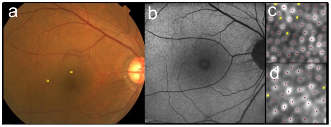
Figure 2. Cone mosaic images of the case with AREDS category 1 (no drusen).

The right eye of 69-year-old male (the same eye as Figure 1) as a representative case with AREDS category 1 (no drusen). The fundus photo (a) did not show any sign of drusen or pigmentary abnormalities. The fundus autofluorescence (FAF) (b) was also unremarkable. After AO image was taken, the 60 pixel by 60 pixel square image was cropped at 2° superior (c) and 5° temporal (d) to the fovea (also shown as yellow squares in a). Cone mosaic was identified automatically at first (red dots in c and d), then added (yellow dots) in manual modification. Cone density were 25,500 cells/mm2 (2,340 cells/deg2) at 2° and 14,030 cells/mm2 (1,290 cells/deg2) at 5°.
