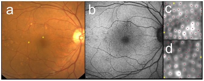
Figure 4. Cone mosaic images of the case with AREDS category 3 (large drusen).

The right eye of 68-year-old female with AREDS category 3. The fundus photo (a) showed large drusen superior and temporal to the fovea. FAF (b) revealed hyper- and hypopigmentation corresponding to the drusen. After AO image was taken, the 60 pixel by 60 pixel square image was cropped at 2° superior (c) and 5° temporal (d) to the fovea (also shown as yellow squares in a). Cone mosaic was identified automatically at first (red dots in c and d), then added (yellow dots) in manual modification. Cone density were 25,700 cells/mm2 (1,990 cells/deg2) at 2° and 14,900 cells/mm2 (1,450 cells/deg2) at 5°.
