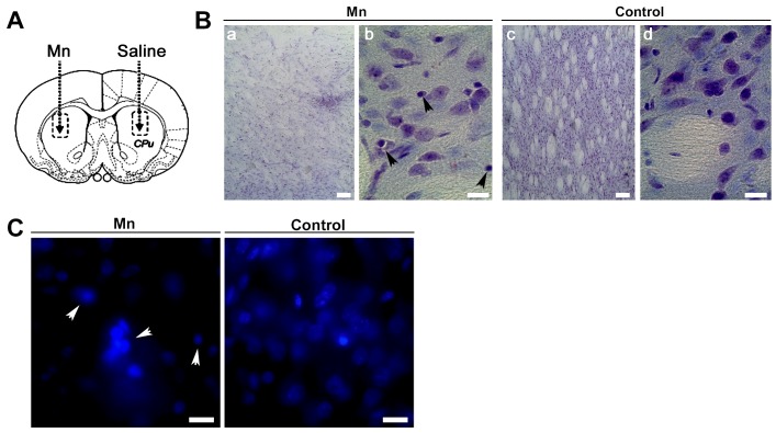
Figure 9.
Mn induced damage into rat striatal tissue (A). Rats were injected into the striatum with 1 µmol Mn (left) and saline solution (right), as indicated with dotted lines. Nissl staining on vibratome brain sections containing striatum from Mn-treated (a,b) and control (c,d) rats (B). Magnification: 10X (a,c) and 40X (b,d); Nuclei staining (C). Arrowheads: cells with shrunken shape and pyknotic nuclei. Magnification: 100X. Samples correspond to sections from rats receiving a single striatum injection of Mn and euthanized 7 days after. Scale bar: 10 µm.

