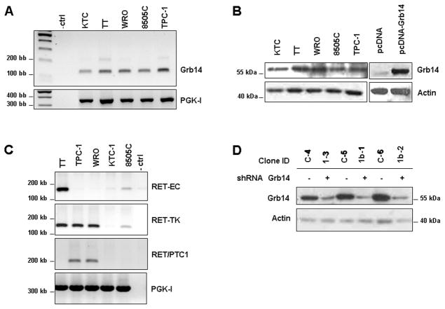
Figure 1. Expression of Grb14 and the RET kinase domain in human thyroid cancer cell lines.
A. RT and real-time quantitative qPCR demonstrate the relative endogenous expression levels of Grb14 in thyroid cancer cell lines. Human PGK-I was used as an internal housekeeping loading control. B. Western blotting comparison of Grb14 protein levels in the same thyroid cancer cell lines. Negative (pcDNA) and positive (Grb14-transfected cells) are shown immediately to the right. Corresponding densitometric values are shown immediately below relative to actin ratios. C. RT-PCR demonstrates expression of the extracellular (EC) and tyrosine kinase (TK) domains of RET. TT cells express full length RET, which yields amplification of both the EC and intracellular TK domains of RET. TPC-1 and WRO cells express the RET/PTC1 rearrangement, which yield amplification of the intracellular TK domain but not the EC domain of RET; instead there is a product using RET/PTC1 primers. Human PGK-I served as an internal housekeeping control. D. Grb14 knockdown in WRO cells was achieved through stable expression of plasmid expressing shRNA targeting the adaptor or scramble sequence controls. Western blotting demonstrates effective Grb14 reduction in 3 independent clones (+) compared to scrambled controls (−).

