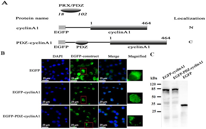
Figure 2. Cytoplasmic localization of a normal nuclear cyclin A1 by fusing with the PDZ domain of L-periaxin.
(A) Schematic representation of cyclin A1 and its chimeric proteins fused with the PDZ domain of L-periaxin. N-terminal hatched boxes indicate EGFP-tagged peptides. The plain numbers on top of the boxes indicate the amino acid residue number of cyclin A1. Italic numbers correspond to the residues flanking the PDZ-domain of L-periaxin. The subcellular localization of each protein, when expressed in RSC96 cells, is indicated on the right. N, nucleus, C, cytoplasm. (B) Fluorescence analysis of RSC96 cells transfected with the plasmids encoding EGFP, EGFP-cyclin A1, and EGFP-PDZ-cyclin A1. Scale bar = 25 μm. (C) GFP fusion proteins are stable in the cell which is shown by Western blotting.

