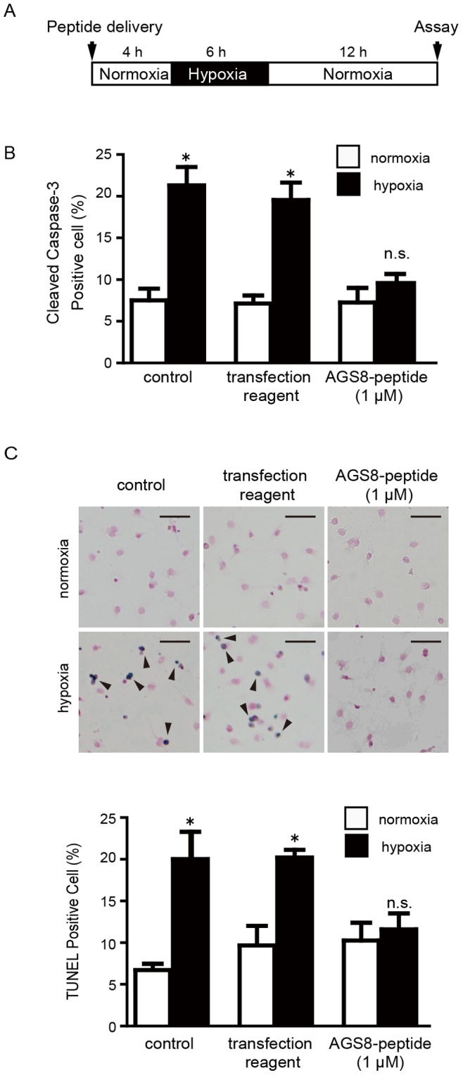
Figure 6. Effect of AGS8 peptide on hypoxia-induced apoptosis of cardiomyocytes.

(A) Neonatal cardiomyocytes were exposed to hypoxia (1% oxygen) or normoxia as indicated duration without or with introduction of AGS8-peptide. Apoptosis was assessed by immunofluorescent detection of the active form of caspase-3 (cleaved caspase-3) (B) or TUNEL stain (C) as described in the experimental procedures. (C) Upper panel indicates representative apoptotic (dark blue, arrow) and non-apoptotic cells (red) after TUNEL staining. Scale bars indicate 100 µm. Approximately 3000 cells of 10 independent fields were counted for each experiments. A separate experiment indicated that AGS8-peptide did not influence the level of AGS8 within the 4-h treatment (1.0 µM peptide; 98.2±7.3% versus no reagent alone group, not statistically significant, n = 4). *, p<0.05 vs cells in normoxia; n.s., not statistically significant. N = 5 from 5 independent experiments.
