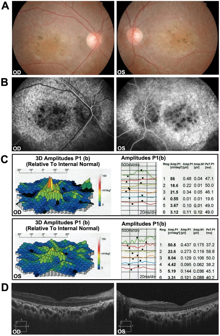
Figure 2. Representative photographs of patients of family 2048.
(A) Fundus photographs showing pigment mottling and yellow-white flecks in both maculae. (B) Fluorescein angiography (FA) images showing the hyperfluorescent flecks extended to the midperipheral retina and fluorescence blocking by the pigment mottling in the mcular. (C) mfERG records showing severe depressed central waveform and significant paracentral/pereferral loss of retinal response. (D) Macular OCTs showing hyper-reflective deposits within the RPE layer and the level of the outer segments of the photoreceptors, thinning of the retinal outer layers and enhanced choroidal reflectivity associated with overlying atrophic retina.

