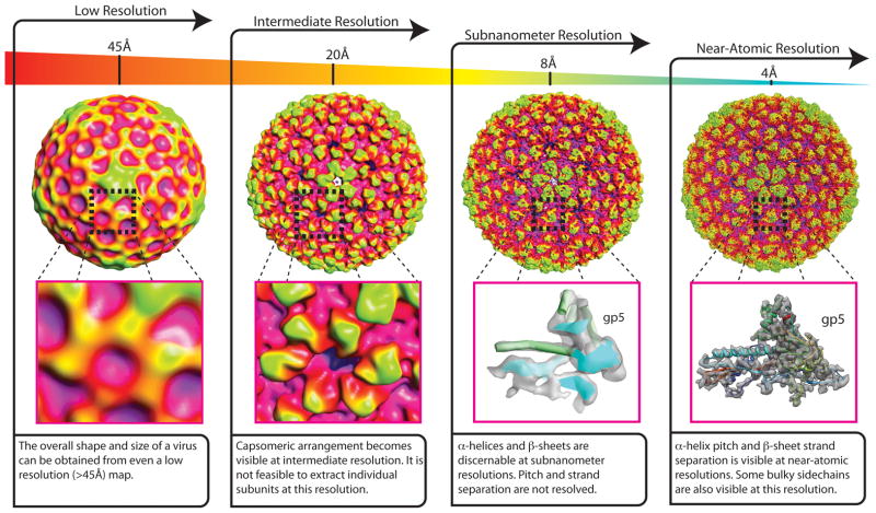
Figure 1.
Features visible as a function of resolution, with Bacteriophage P22 Procapsid shown for reference [EMD-1824]. At low-resolutions (~45Å) very basic information about the shape and size of a virus are apparent in the 3-D reconstruction. As the resolution of the reconstruction increases to near 20Å, it becomes possible to identify the arrangement of capsomeres in the capsid shell. At subnanometer resolutions (<10Å), α-helices and β-sheets become visible. As the resolution of a reconstruction pushes towards near-atomic resolutions, α-helix pitch and β-sheet strand separation are apparent. Additionally it is possible to visualize bulky side-chains in the protein subunits that comprise the map.

