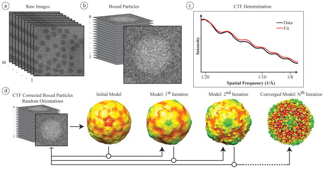
Figure 3.
Generalized schematic for single particle reconstruction. (a) A single cryo-EM micrograph or CCD frame can contain anywhere from just a few to hundreds of particles. (b) The first step in reconstruction is to box the individual particles from all micrographs. Depending upon the software used, it may be necessary to invert the particles contrast prior to alignment and reconstruction. (c) Once boxed, the power spectrum of the boxed particles of a micrograph can be used to determine the particles’ CTF. (d) Using the determined CTF parameters, the particles can be aligned to a model and then reconstructed to a better-resolved map. The first step in this process is to build an initial model, as discussed in text. While there are a variety of different alignment schemes used in single particle virus reconstruction, the general approach is to align the single particle images to references projected from either an initial model or a previously reconstructed model, and then iteratively refine this model until it converges.

