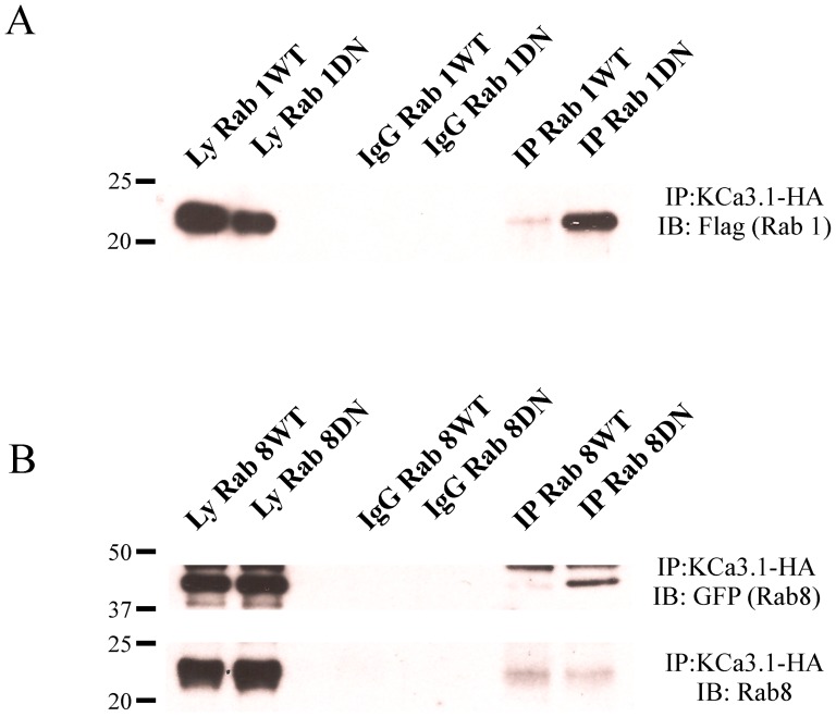
Figure 5. KCa3.1 immunoprecipitates with Rab1 and Rab8.
A. Co-IP of HA-tagged KCa3.1 and either WT or DN Rab1 (Flag) was carried out in HEK293 cells as described in the Methods. Total cell lysates were subject to IP using either an α-HA Ab (lanes 5, 6) or an α-V5 Ab as IgG control (lanes 3, 4) and subsequently blotted for Rab1 (α-Flag). B. Co-IP of HA-tagged KCa3.1 and either endogenous (bottom panel) or GFP-tagged WT and DN Rab8 (top panel). Total cell lysates were subject to IP as in A and subsequently IB for either endogenous Rab8 (α-Rab8) or GFP-tagged Rab8 (α-GFP). Lysates for WT and DN Rab1 or 8 are shown in the first two lanes of each blot (5 μg loaded per lane). These data confirm an association between KCa3.1 and Rabs1 and 8. Data are representative of 3 experiments.

