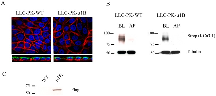
Figure 8. Basolateral targeting of KCa3.1 is independent of the adaptor protein, μ1B.
A. BLAP-KCa3.1 was transduced in to either WT LLC-PK1 cells (left panels) or LLC-PK1 cells stably expressing μ1B (right panels) grown to confluence on Transwell® filters. The AP and BL membranes were biotinylated using recombinant BirA and biotinylated proteins labeled with streptavidin-Alexa555 (red). Apical membrane was co-labeled with WGA-Alexa488 (green). Nuclei were labeled with DAPI (blue). The top panels show a single confocal section through the mid-plane of the cells and the bottom panels show a z-stack. KCa3.1 was detected exclusively in the BL of both LLC-PK1 clones. B. Either AP or BL membranes of WT LLC-PK1 cells (left panel) or LLC-PK1 cells stably expressing μ1B (right panel) grown to confluence on Transwell® filters were biotinylated using BirA followed by streptavidin labeling and subsequent IB to determine localization of KCa3.1. KCa3.1 was localized specifically to the BL membrane in both LLC-PK1 clones. Tubulin was used as a loading control. Blots are representative of 3 separate experiments. 20 μg of protein was loaded per lane. C. IB confirming expression of Flag-tagged μ1B in the LLC-PK1-μ1B cell line.

