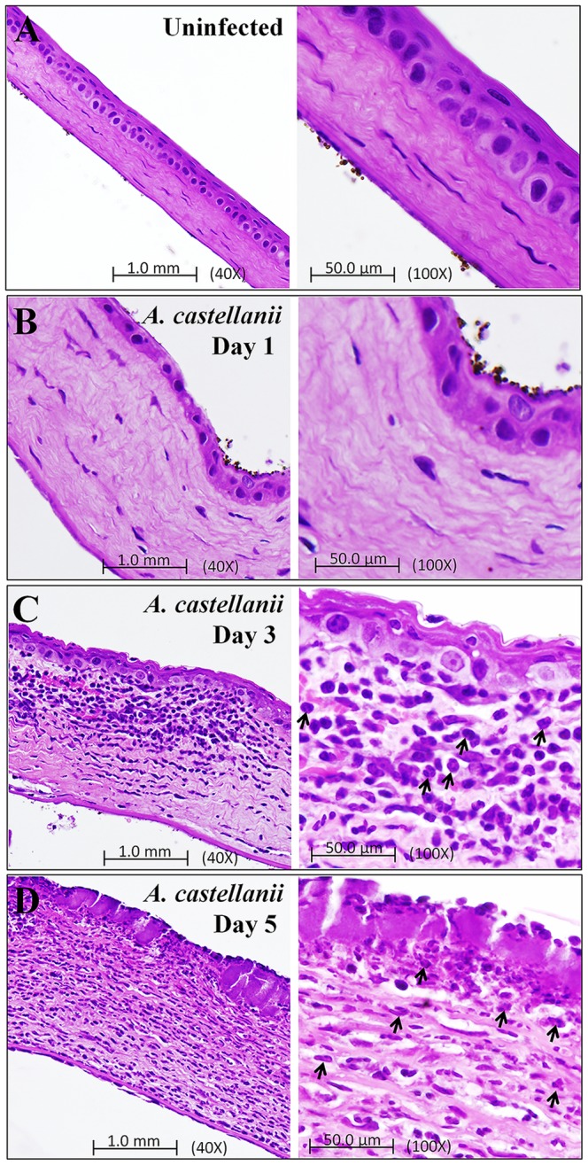
Figure 8. Pathological evaluation of A. castellanii induced inflammation in Chinese hamster corneas.

Photomicrograph of corneas from a Chinese hamster infected with A. castellanii trophozoites-laden contact lenses. Animals were anesthetized and sacrificed on 1, 3, and 5 days postinfection. The histopathological features of the uninfected and infected corneas included - (8A) Cornea from control-uninfected animal was normal with regular pattern of corneal epithelium, stroma, and corneal endothelium. (8B) Infected corneal section of day 1 postinfection shows very mild inflammation in stroma as compared to uninfected corneal section. (8C and 8D) Infected corneal section of 3 and 5 days postinfection show epithelial ulceration, focal thickening, PMNs infiltration, corneal thickening, marked lamellar connective tissue disruption, and extensive stroma swelling when compared with uninfected corneal section. PMN cells (Arrows) were present in the stroma. (8A-8D) Left panel shows 40X magnification and right panel shows 100X magnification.
