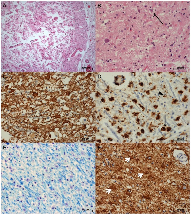
Figure 2. Examples of histopathologic findings.
A: Pannecrosis, parietal lobe. HE stain. B: Neuronal necrosis (arrow), frontal lobe. HE stain. C: glial scarring of the white matter. GFAP stain. D: Inflammation of the white matter (black arrows: macrophages, black arrow head: microglia). CD68 stain. E: Patchy loss of myelin structure in the occipital lobe. Klüver-Barrera stain. F: Astrogliosis of the putamen (white arrow heads: astrozytes). GFAP stain.

