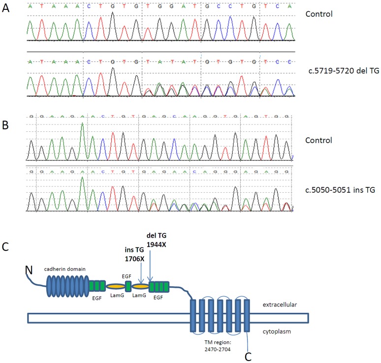
Figure 1. TG dinucleotide repeats variants in spina bifida.
A: Sequence trace of control (top) and C.5719–5720delTG (bottom). B: Sequence trace of control (top) and TG duplication (bottom). C: Schematic representation of the CELSR1 predicted protein structure (accession number Q9NYQ6) with the domains and approximate position of TG repeats variants.

