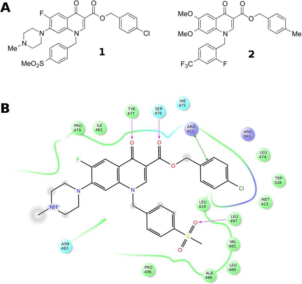
Figure 1.
A) Chemical structures of 1 and 2 as representative quinolones TSII-NNIs. B) Schematic representation of the interaction between NS5B polymerase and compound 1 (PDB ID 3PHE). NS5B residues lying within a distance of 4 Å from the bound ligand are shown and color coded as follows: green-hydrophobic; purple-basic; cyan-polar. The TSII pocket is displayed with a line, exhibiting the color of the nearest protein residue. The gap in the line shows the opening of the pocket. Specific interactions between ligand atoms and protein residues are marked with lines: pink, H-bonds to protein backbone; green, π-π stacking interactions. Ligand atoms that are exposed to solvent are marked with gray spheres.

