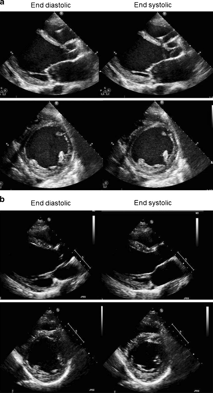
Fig. 1.
a Initial phase (week 1). Transthoracic echocardiographic images representative of case 1; diastolic (left) and systolic (right) still frames from parasternal long axis, short axis views. Side box. LVEDD 68 mm. LVESD 62 mm. Estimated LVEF 10 %. b After ECMO and heart failure medical treatment (6 months). Transthoracic echocardiographic images representative of case 1; diastolic (left) and systolic (right) still frames from parasternal long axis, short axis views. Side box. LVEDD 65 mm. LVESD 44 mm. Estimated LVEF 35–40 %

