
Figure 5. Mechanisms of penumbra pathology.
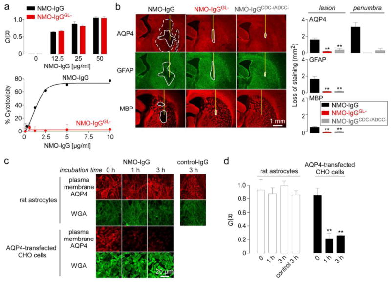
a. (Top) Binding of control and deglycosylated NMO-IgG (NMO-IgGGL-) to AQP4-expressing CHO cells showing red-to-green fluorescence ratio (R/G) at indicated NMO-IgG concentrations. (Bottom) Cytotoxicity (by Alamar blue assay) following incubation for 1 h with NMO-IgG or NMO-IgGGL- and 5% complement (S.E., n=3). b. AQP4, GFAP and MBP immunofluorescence at 5 days after injection with NMO-IgG, NMO-IgGGL- or NMO-IgGCDC-/ADCC-. (Right) Summary of lesion areas (S.E., n=3, ** P < 0.01). c. Internalization of AQP4 in rat primary astrocyte cultures. Cells were incubated for 1 or 3 h at 37 °C with 100 μg/ml NMO-IgG or control IgG. Surface AQP4 immunofluorescence shown in red, with plasma membrane marker (WGA) in green. AQP4 internalization in AQP4-expressing CHO cells shown as positive control. d. R/G ratios from experiments as in c (S.E., n=3, ** P < 0.01).