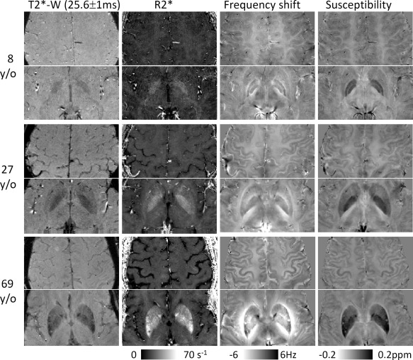
Figure 1.

Images derived from multiecho gradient echo MRI: T2*‐weighted magnitude (T2*‐W), R2*, frequency shift, and susceptibility. Magnitude and R2* show good contrast between iron‐rich nuclei and surrounding tissues but show very weak contrast between gray and white matter. In comparison, both phase and susceptibility show not only good contrast between gray and white matter but also between iron‐rich nuclei and surrounding tissues. Although magnitude, R2*, and susceptibility are all well‐localized contrasts with respect to the anatomical structures, frequency shift is affected by surrounding susceptibility distributions. As shown by the color bar, both phase and susceptibility are displayed in a reversed scale so that brighter intensity corresponds to more diamagnetic susceptibility.
