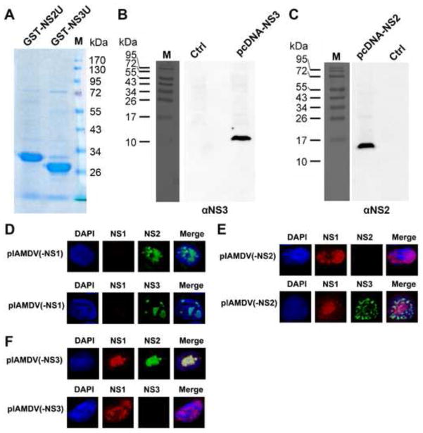
Fig. 3. Analyses of the specificity of the rat anti-NS2 and anti-NS3 antibodies.
(A) Coomassie blue staining of the purified GST-NS2U and GST-NS3U. ~2 μg of the purified GST-NS2U and GST-NS3U proteins were resolved in SDS-10%PAGE gel. The gel was stained using Coomassie blue. A molecule weight marker is shown. (B&C) Western blot analysis of NS2 and NS3 expression. CrFK cells were transfected with constructs pcDNA-NS2, pcDNA-NS3, and pcDNA3 (as a control, ctrl). At 2 days post-transfection, transfected cells were harvested, lysed and resolved in SDS-15%PAGE (panel B) or SDS-12%PAGE (panel C) followed by Western blot analysis using the rat anti-NS3 or anti-NS2 antibody. (D–F) Immunofluorescence analysis of NS expression. CrFK cells were transfected with pIAMDV(-NS1) (D), pIAMDV(-NS2) (E) and pIAMDV(-NS3) (F). At 4 days post-transfection, the transfected cells were analyzed by immunofluorescence using rabbit anti-NS1 and rat anti-NS2 antibodies or rabbit anti-NS1 and rat anti-NS3 antibodies, as indicated. Confocal images were taken at × 60 magnification (objective lens). Nuclei were stained with DAPI.

