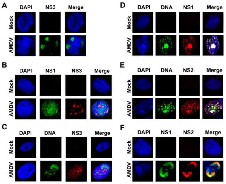
Fig. 4. Immunofluorescence analysis of AMDV NS2 and NS3 expression.
CrFK cells were infected with AMDV-G at an MOI of 1 or mock-infected. (A–C) Analysis of NS3 expression. At 4 days p.i., cells were stained with rat anti-NS3 (A), rabbit anti-NS1 and rat anti-NS3 (B), respectively. The cells were also analyzed by FISH-Immunofluorescence with a biotintylated AMDV-G DNA probe and a rabbit anti-NS3 antibody (C). (D–F) Analysis of NS2 expression. At 4 days p.i., cells were analyzed by FISH-Immunofluorescence with a biotintylated AMDV-G DNA probe and a rabbit anti-NS1 antibody (D) or a rat anti-NS2 antibody (E). The cells were also co-stained with rabbit anti-NS1 and rat anti-NS2 antibodies (F).
Confocal images were taken at × 60 magnification (objective lens). Nuclei were stained with DAPI.

