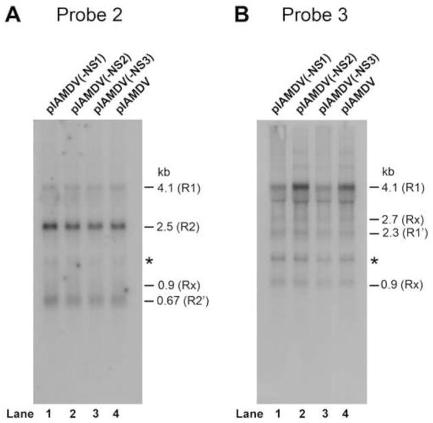
Fig. 5. Northern blot analysis of AMDV mRNAs in the cytoplasm of the cells transfected with pIAMDV and the NS knockout mutants.
CrFK cells were transfected with pIAMDV, pIAMDV(-NS1), pIAMDV(-NS2) and pIAMDV(-NS3), respectively. At 4 days post-transfection, cytoplasmic RNA was extracted from transfected cells and analyzed by Northern blot using Probe 2 and Probe 3, which span a region of nt 180-1600 and nt 1121-1960, respectively, of the AMDV-G genome (diagramed in Fig. 1A). The identity and the size of detected viral mRNAs, which have been confirmed previously (Qiu et al., 2006a), are shown to the right of each panel.

