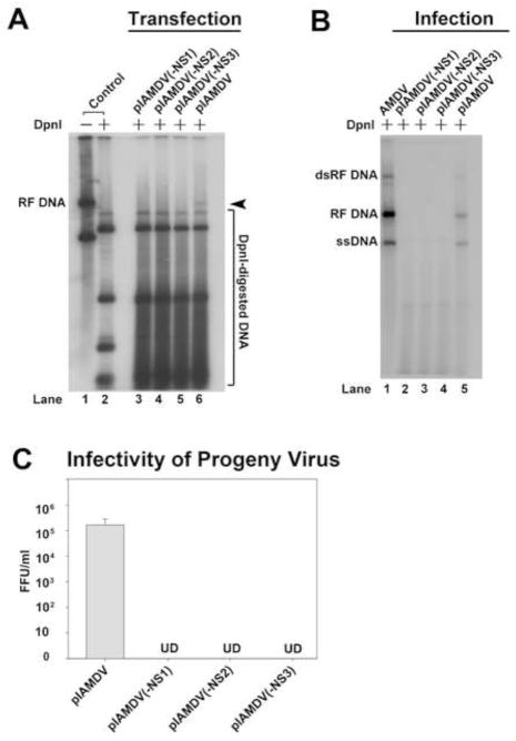
Fig. 6. Analyses of the NS knockout mutant in the context of an AMDV infectious clone.
(A&B) Southern blot analysis of viral DNA replication. (A) CrFK cells were transfected with pIAMDV, pIAMDV(-NS1), pIAMDV(-NS2) and pIAMDV(-NS3), respectively. At 6 days post-transfection, transfected cells were collected and used to prepare Hirt DNA. (B) CrFK cells were infected with virus samples prepared from the above CrFK cells transfected with pIAMDV, or pIAMDV(-NS1), pIAMDV(-NS2), and pIAMDV(-NS3). At 6 days p.i., infected cells were collected and used to prepare Hirt DNA samples. The Hirt DNA samples were digested with DpnI for Southern blotting. The blots were probed with a full-length AMDV probe (Qiu et al., 2006a). The identity of major bands on the blots is indicated. RF DNA, replicative form viral genome; dRF DNA, double replicative form viral genome; ssDNA, single-stranded viral genome. Cells infected with AMDV-G were used a control (AMDV). (C) Titration of virus production in cells infected with progeny virus generated from transfection. CrFK cells were infected with virus samples prepared from the above CrFK cells transfected with pIAMDV, or pIAMDV(-NS1), pIAMDV(-NS2) and pIAMDV(-NS3), and were harvested at 6 days post-transfection. The cells were frozen and thawed three times, and briefly centrifuged. The supernatant was collected, serially diluted, and used to infect fresh CrFK cells in order to titrate FFU. Results shown represent the averages and standard deviation from at least three independent experiments. UD, undetectable.

