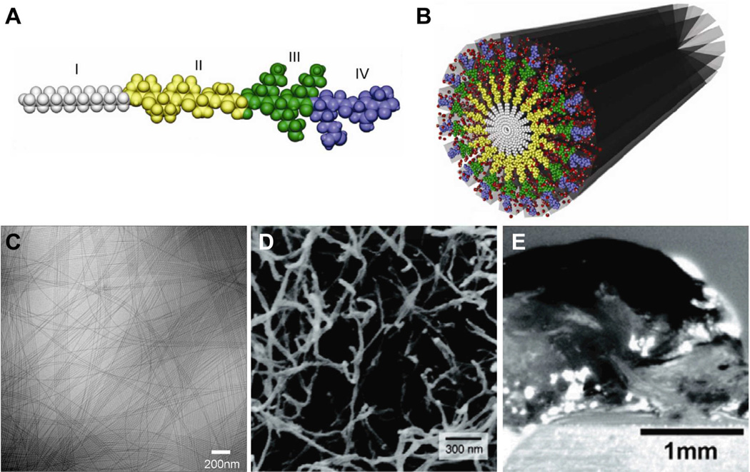
Fig. 2.
(A) Molecular graphics representation of a canonical peptide amphiphile (PA) molecule, showing the (I) hydrophobic alkyl tail, (II) β-sheet forming residues, (III) charged residues, and (IV) bioactive peptide sequence. (B) Molecular graphics representation of a cylindrical nanofiber formed by peptide amphiphiles in water. (C) Cryogenic TEM of PA nanofibers, (D) SEM micrograph of a nanofiber network, and (E) optical image of a hydrogel formed by the nanofiber network. Adapted with permission from reference 36. Copyright 2011 Elsevier Ltd.

