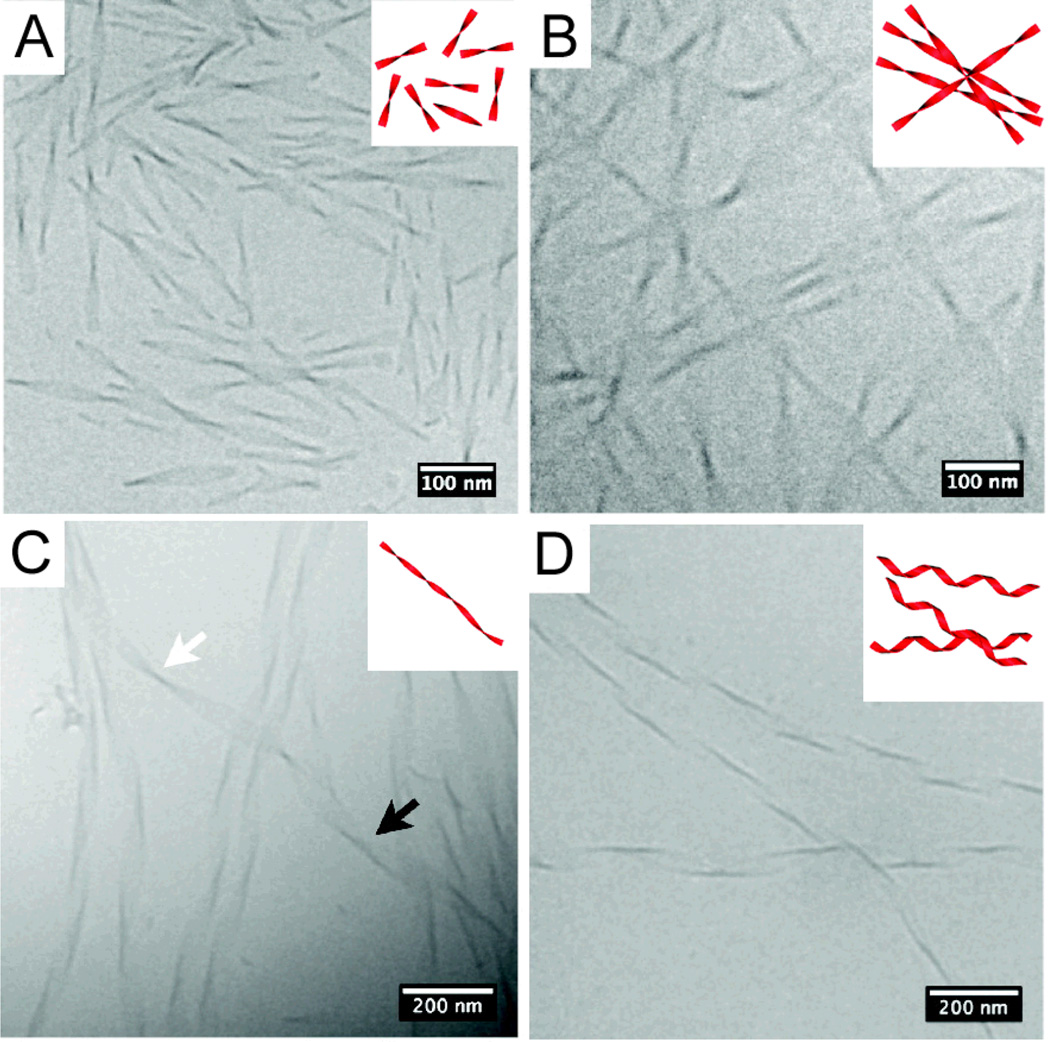
Fig. 4.
Chemical structure of a series of amphiphilic peptide amphiphiles (PAs) with different numbers of dimeric repeats of hydrophobic (valine) and hydrophilic (glutamic acid) amino acids (top). Transmission electron micrographs of ribbon-like assemblies of the PAs containing two, four, and six dimeric units (middle), and their respective molecular graphics representations. As the number of dimeric repeats is increased from two to six, ribbon twisting increases and lateral width of the assemblies is reduced from 100 nm down to 10 nm. Adapted with permission from reference 80. Copyright 2013 American Chemical Society.

