Figure 1.

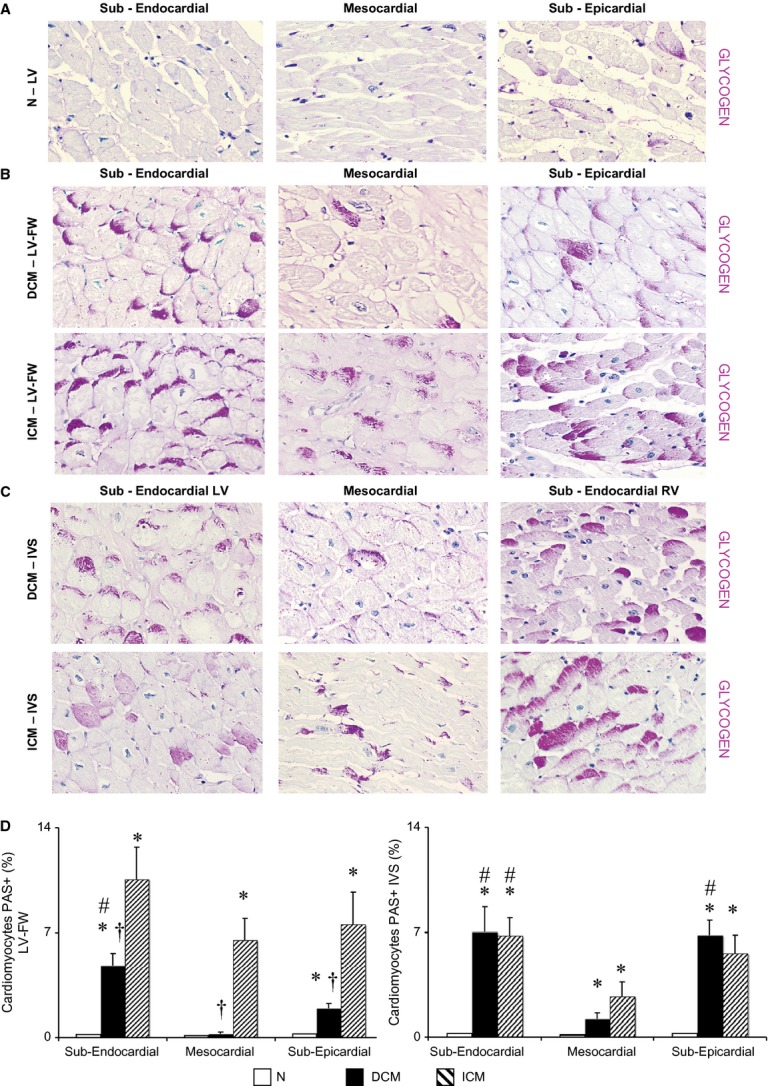
Extent and distribution of regional left ventricle (LV) glycogen deposits as shown by periodic acid-Schiff (PAS) staining on histological sections. (A–C) Representative images of PAS stained sections for each myocardial layer in the LV-free wall (FW) of normal donor (N) hearts, in the LV-FW and in the inter-ventricular septum (IVS) of either dilated cardiomyopathy (DCM) and ischemic cardiomyopathy (ICM) hearts (magnification 400 × ); (D) Intracellular glycogen amount in LV-FW and IVS of N (n = 8), of DCM (n = 11) and ICM (n = 12) hearts. Values are means ± SEM. *P < 0.05 versus Normal, # versus mesocardial layer, †P < 0.05 versus corresponding layer of ICM heart.
