
Figure 1.
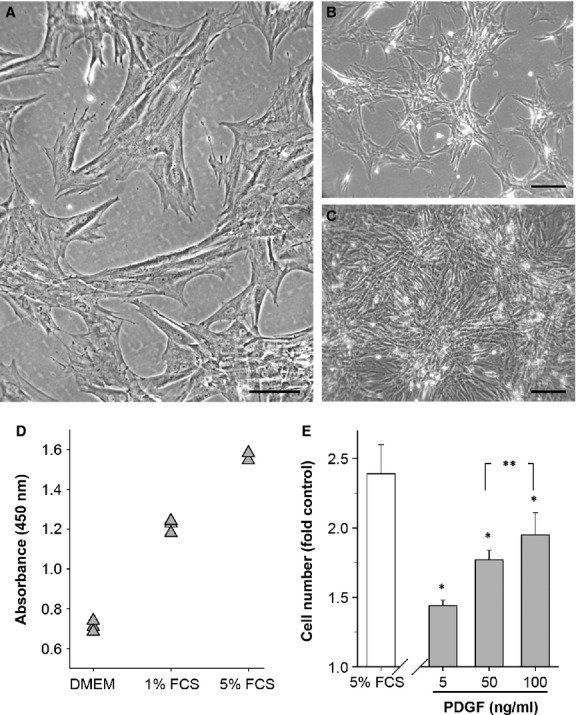
A culture model of proliferating adult colonic circular smooth muscle cells (CSMC) shows the mitogenic role of platelet-derived growth factor (PDGF)-BB. (A–C) Phase contrast images showing typical appearance of CSMC at passage 2 in vitro after plating (A), during growth (B) and at confluence (C). Scale bars: A, 100 μm; B and C, 200 μm. (D) Typical experimental outcome of CSMC growth after 48 hrs in either medium alone (DMEM) or DMEM + FCS using the Wst-8 assay. OD450 nm is proportional to cell number, shown here for triplicate wells per condition. (E) Average growth response of CSMC relative to serum-free control in either 5% FCS or PDGF-BB. All values significantly greater than control (P < 0.05). Cell number is expressed as the ratio of WST-8 reaction product in treated versus control wells, for FCS (n = 8) or PDGF-BB (n = 4), with triplicate wells per experiment. *P < 0.05 versus control; **P > 0.05.