
Figure 5.
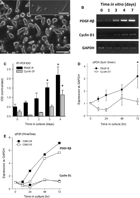
The appearance of mRNA for platelet-derived growth factor (PDGF)-Rβ in freshly isolated adult circular smooth muscle cells (CSMC) precedes entry into cell cycle. (A) Appearance of CSMC at 6 hrs after isolation, showing transition from a long, bipolar appearance to a rounded, surface-attached appearance. Phase contrast main image (scale bar, 100 μm) and inset, Nomarski optics (scale bar, 80 μm). (B) Representative image of RT-PCR experiment showing appearance of mRNA for PDGF-Rβ occurs before that of cyclin-D1. (C) Quantification of RT-PCR showing time course of onset of expression of PDGF-Rβ in freshly isolated CSMC from control rats, with outcome normalized to GAPDH. CSMC initially lack significant expression of PDGF-Rβ, which first appeared by Day 3 and increased thereafter (*P < 0.05 versus control). (D) Quantitative PCR showing that the appearance of mRNA for PDGF-Rβ precedes mRNA for cyclin D1, a marker of proliferation. Data are the means of three experiments (i.e., cells from three separate animals) with two replicate wells per time-point (*P < 0.05 versus control). (E) Outcome of two independent experiments showing time course of up-regulation of mRNA for PDGF-Rβ, which consistently preceded that of cyclin D1. Data are the average of two wells per condition per animal, assessed by qPCR using PrimeTime probes.