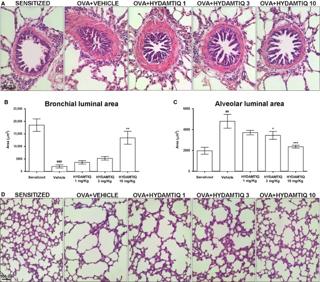
Figure 3.
Evaluation of bronchiolar and alveolar luminal area in guinea pig lungs. The luminal area of bronchioles (A and B) and alveoli (C and D) were evaluated in haematoxylin-and eosin-stained sections and quantified by morphometric analyses. Data are means ± SEM. ##P < 0.01 and ###P < 0.001 versus Sensitized; *P < 0.05, **P < 0.01 and ***P < 0.001 versus Vehicle.

