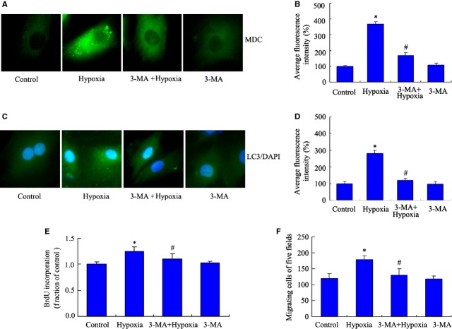
Figure 3.
3-MA inhibits autophagy and decreases the proliferation of pulmonary arterial smooth muscle cells (PASMCs) induced by hypoxia. PASMCs were pre-incubated with 3-MA (5 mM) for 30 min. after 24 hrs, cells were exposed to hypoxia and normoxia chamber for 24 hrs. (A) The formations of autophagic vacuoles were detected by punctated monodansylcadaverine (MDC) immunofluorescence staining. Microphotographs are shown as representative results from three independent experiments. Images are at 1000×. (B) The corresponding linear diagram of MDC staining results. (C) PASMCs were processed for LC3 immunofluorescence staining. (D) The corresponding linear diagram of LC3 staining. (E) Cell proliferation was measured by 5-bromo-2′-deoxyuridine (BrdU) assay. n = 5, mean ± SD. *P < 0.05 versus control group, #P < 0.05 versus hypoxia group. (F) Migration of PASMCs exposed to 3-MA under hypoxia was detected by transwell assay. n = 5, mean ± SD. *P < 0.05 versus control group, #P < 0.05 versus hypoxia group.

