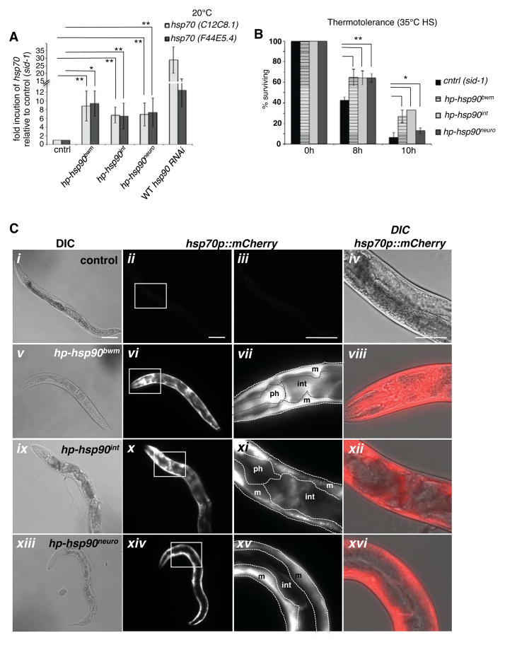
Figure 4. Tissue-specific knockdown of hsp90 cell-non-autonomously induces the HSR.
(A) Bodywall-muscle, intestine- and neuron-specific hsp90 RNAi induces basal levels of hsp70 (C12C8.1 and F44E5.4) expression at 20°C, compared to control animals (sid-1). Wild type animals allow import of dsRNA from surrounding tissues, leading to higher induction of organismal hsp70 than in the tissue-specific knockdown lines. Bargraphs represent combined mean values of three independent experiments (means ± s.e.m.) **P-value < 0.05. (B) Thermosensitivity of young adult animals (n = 100) expressing the indicated tissue-specific hp-hsp90 construct exposed to 35 °C heat stress. Bargraphs represent combined mean values of three independent experiments (means ± s.e.m.) **P-value < 0.01. *P-value < 0.05. (C) Tissue-specific knockdown of hsp90 induces expression of the hsp70 reporter (hsp70p::mCherry) at 20°C. DIC images of synchronized young adult (i) control animals (sid-1), (v) hp-hsp90bwm, (ix) hp-hsp90int, and (xiii) hp-hsp90neuro animals expressing the hsp70 reporter. Expression of the hsp70p::mCherry in (ii) sid-1 control animals, (vi) hp-hsp90bwm, (x) hp-hsp90int, and (xiv) hp-hsp90neuro. (iii, vii, xi, xv) 20x magnification of control (iii) and tissue-specific hsp90 knockdown lines (vii, xi, xv) indicating expression of hsp70p::mCherry in the pharynx (ph), intestine (int) and bodywall muscle (m). (iv, viii, xii, xvi) Overlay of DIC Nomarski and hsp70p::mCherry (red). Scalebars = 100 μm. See also Figure S4.

