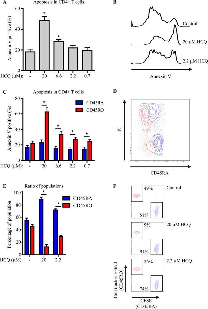
Figure 2. HCQ treatment preferentially induces apoptosis in CD4+CD45RA-CD45RO+ T cells.
(A) Apoptosis (by Annexin V staining) in human CD4+ cells, cultured in the presence of HCQ, n=8. (B) Histograms of (A). (C) Apoptosis in human CD4+CD45RA+CD45RO- and CD4+CD45RA-CD45RO+ cells, n=7. (D) FACS-plot of HCQ treated CD4+CD45RA+CD45RO- (blue) and CD4+CD45RA-CD45RO+ (red) cells stained with propidium iodide (PI). (E+F) Sorted CD4+CD45RA+ (CFSE) or CD4+CD45RO+ (EF670) cells were mixed at a 1:1 ratio and cultured in the presence of HCQ, (n=4).

