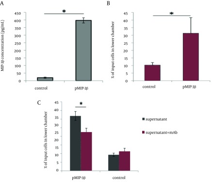
Figure 1. Expression and Chemotactic Activity of MIP-3beta.

A) Sandwich ELISA was used to confirm the expression of MIP-3beta. pcDNA 3.1+ was used as control in transfection experiments. B) Induced chemotaxis of CCR7+ HUT-78 cell line by the secreted MIP-3beta was evaluated by examining the percentage of migrated cells to the lower chamber in the presence of pMIP-3beta-transfected cell supernatant. Backbone plasmid-transfected supernatant was used as control. C) Inhibition of MIP-3beta induced chemoattraction by antibody-mediated neutralization of chemokine (mAb: MIP-3beta neutralizing monoclonal antibody). All experiments were performed in duplicate and repeated three times. Results are expressed as mean percentage of migrated cells to the lower chamber of transwell ± standard deviation (SD). Asterisks represent significant deference (p < 0.01)
