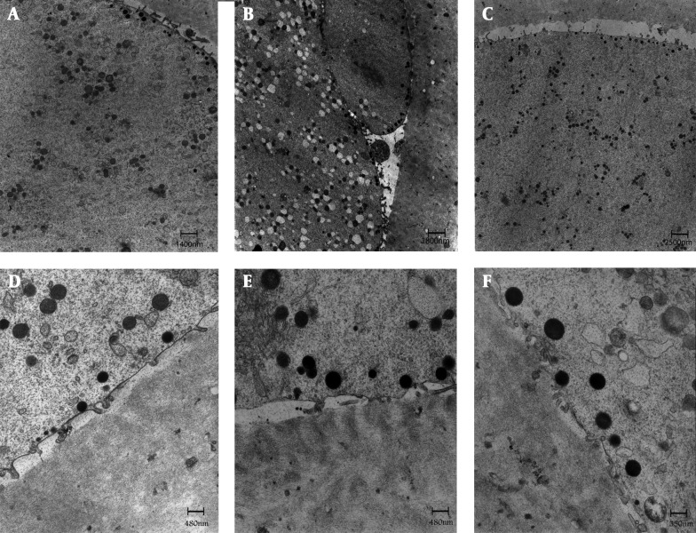
Figure 1. General Fine Structure and Organelle Microtopography are shown by Transmission Electron Microscopy.
In control (A, D), GV (B, E) & MI (C, F) stage oocyte after IVM. Microvilli are numerous and long on the oolemma of oocytes. A rim of electron-dense cortical granules (arrows) is seen just beneath the oolemma of oocytes. ZP = zona pellucida; mv = microvilli; CG = cortical granules; O = oocyte; PVS = perivitelline space; PB = polar body

