Figure 3.

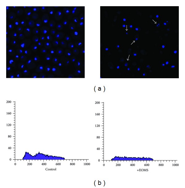
Laser scanning confocal microscopy after Hoechst staining and cell cycle analysis by FACS. C. albicans cells untreated or treated with EOMS were observed by a LSCM after Hoechst staining (a) and analysis by FACS of cell cycle (b).

Laser scanning confocal microscopy after Hoechst staining and cell cycle analysis by FACS. C. albicans cells untreated or treated with EOMS were observed by a LSCM after Hoechst staining (a) and analysis by FACS of cell cycle (b).