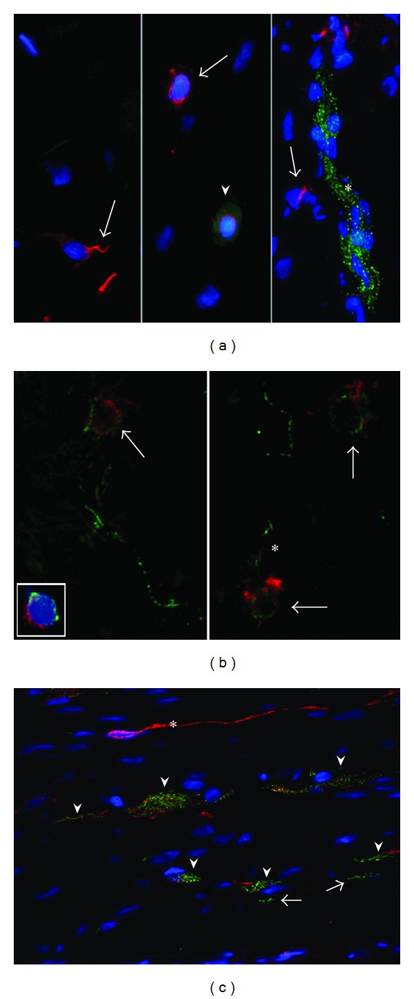
Figure 4.

Double staining of N-cadherin and C-kit. (a) Arrows show cells in the bladder expressing C-kit (arrows, red). They lack expression of N-cadherin (green). Arrowhead shows a round cell with perinuclear expression of C-kit and diffuse cytoplasmic background signal for N-cadherin, highly resembling a mast cell. Elongated clusters of punctate N-cadherin+ cells lack expression of C-kit (asterisk). Magnification ×630. (b) Staining lacking DAPI for better orientation. Arrows show cells in the bladder expressing C-kit and punctate N-cadherin. These cells seem to give rise to N-cadherin+ branches (asterisk). Magnification ×1000. (c) N-cadherin+/C-kit+ cells running parallely (arrowheads) are neighboured by a slender elongated N-cadherin−/C-kit+ cell body (asterisk) and several smaller N-cadherin+/C-kit− cells (arrows) in the jejunum. Magnification ×630.
