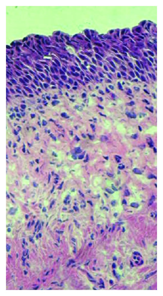
Figure 6.

Representative hematoxylin and eosin stain of urothelial area of the specimens used. Normal urothelium consists of approximately 3–5 cell layers. Note that no abnormal thickening of the urothelial layer or abnormal suburothelial morphology is found.
