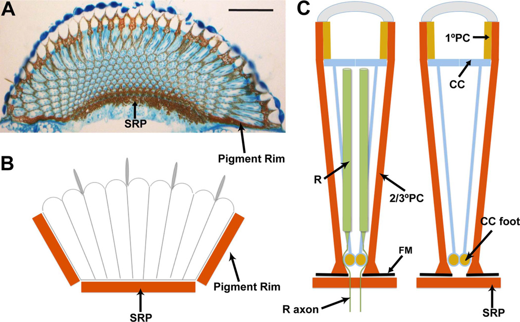
Figure 1.
Summary of the screening pigments in the Drosophila eye. A: A longitudinal section through an eye stained with toluidine blue. Arrows point to the subretinal pigment layer (SRP) and the pigment rim. B: Schematic image of a longitudinal section showing how the eye sits in a pigmented cup made basally by the subretinal pigment, and on the sides by the pigment rim. C: Schematic summary of the screening pigments of individual ommatidia. Two images are shown; the one to the right has the photoreceptors (R) removed to allow a clear view of how the cone cells (CC) send fibers to the base, where ommochrome pigment accumulates in their feet. The primary pigment cells (1°PC) surround the lens unit. The secondary and tertiary pigment cells (2/3°PC) extend the depth of the eye and end basally in enlarged feet that sit directly on the fenestrated membrane (FM). The photoreceptors extend much of the retina and then send axons through a hole in the fenestrated membrane. Immediately below the fenestrated membrane lies the subretinal pigment layer (SRP). Scale bar = 50 µm in A. [Color figure can be viewed in the online issue, which is available at wileyonlinelibrary.com.]

