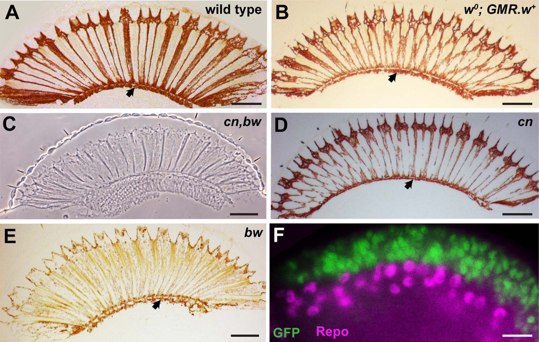
Figure 2.
A–E: The presence of the subretinal pigment layer in various genetic backgrounds that manipulate pigment expression. A: A brightfield image through a section of a wild-type eye shows the characteristic subretinal pigment layer lying directly beneath the retina (arrowhead). B: A brightfield image of a white mutant eye (w0) in the presence of a transgene driving expression of w+ in the presumptive retina (GMR-w+), which creates a normally pigmented eye, with a normal subretinal pigment layer (arrowhead). C: A phase contrast image of a section through a cn,bw mutant eye in which pigment is absent from the entire eye including the subretinal region. D: A brightfield image of a section through a cn mutant eye in which a general reduction in pigment levels in the eye is seen, and a reduced subretinal pigment layer is still present (arrowhead). E: A brightfield image of a section through a bw mutant eye in which a general reduction in pigment levels in the eye is seen, and a reduced subretinal pigment layer is still present (arrowhead). F: Fluorescence microscopy image of a late third-instar GMR-Gal4; UAS-GFP eye disc stained for GFP (green) and Repo (magenta) a pan-glia marker. The GMR-expressing cells overlie the glia, and there is no overlap in expression. The image is taken from the region of the disc that rolls laterally and allows the upper and lower layers of the tissue to be viewed in cross section. Scale bar = 50 µm in A–E; 10 µm in F.

