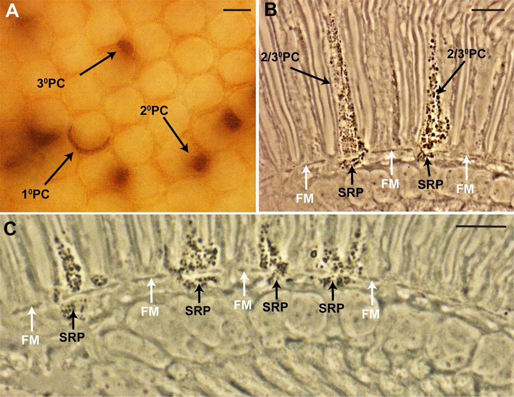
Figure 3.
The presence of pigment in the secondary/tertiary pigment cells corresponds with pigment in the underlying subretinal pigment layer. All images are from flies of the genotype w0; tub>y+>Gal4, UAS-w+ heat shocked in the first 2 days of pupation to produce a random array of cells expressing pigment. A: Cross-sectional brightfield image from a whole mount eye (before embedding for sectioning) focused to the level of the lenses. A random array of pigment cell types is evident. Primary pigment cells (1°PC) appear as crescent shaped, and surround half of a lens unit. The secondary pigment cells (2°PC) are located between two adjacent ommatidia, whereas the tertiary pigment cells (3°PC) are located at the vertices between three ommatidia. B: Longitudinal section through an eye similar to the one shown in A viewed with phase contrast microscopy. Pigment is evident as dark granules. Two cells of the secondary/tertiary pigment cell class (2/3°PC) express pigment, and each has a corresponding patch of subretinal pigment (SRP) beneath the fenestrated membrane (FM; white arrows). C: An image similar to B, but enlarged in the basal region to show that whenever subretinal pigment is present there is pigment in the overlying pigment cells of the retina. Scale bar = 10 µm in A–C. [Color figure can be viewed in the online issue, which is available at wileyonlinelibrary.com.]

