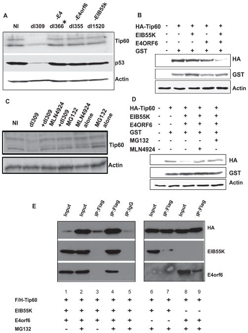
Figure 2. Adenovirus oncoproteins EIB55K and E4orf6 promote proteasome-dependent Tip60 degradation.
(A) MCF10A cells were infected with different strains of adenovirus and lysates prepared 72 hours after infection were probed with indicated antibodies.
(B) U2OS cells were transformed with indicated plasmids. Cells were harvested 48 hours after transfection and lysates immunoblotted with indicated antibodies. GST-expressing plasmid was cotransfected as a transfection control.
(C) Inhibition of the proteasome or Cullins activity stabilizes Tip60 in dl309-infected cells. MCF10A cells were infected by dl309 strain and MG132 or MLN4924 was added to infected cells at 4 or 24 hours before harvesting (72hrs post infection), respectively. Lysates were probed with indicated antibodies.
(D) U2OS cells were transfected with indicated plasmids and addition of either MG132 or MLN4924 stabilizes the Tip60 protein. Rest is as in (B).
(E) Interaction between Flag/HA-Tip60, EIB55K and E4orf6. U2OS cells were transfected with indicated plasmids for 48 hours and treated for 5 hours with or without MG132 before harvest. Anti-flag antibodies and control IgG were used for immunoprecipitation and immunoprecipitated proteins resolved and probed with indicated antibodies. Immunoblot shows co-precipitation of Flag/HA-Tip60, EIB55K, and E4orf6. Input lanes contain 5% of lysates input into the immunoprecipitates.

