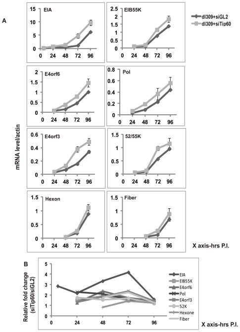
Figure 3. Tip60 represses expression of viral transcription factor EIA.
(A) Levels of mRNA of different viral genes; EIA (early), EIB55K (early), E4orf6 (early), Pol (early), E4orf3 (early), 52/55K (early/late) Hexone (late) and Fiber (late) were measured at indicated time points after viral infection by RT-qPCR and normalized to β actin. MCF10A cells transfected with siGL2 or siTip60 were infected with dl309. Mean ± SD (n=3).
(B) Relative expression of different viral genes in siTip60 transfected cells relative to siGL2 transfected cells. EIA mRNA shows most enhancement at 72 hr after infection in cells where Tip60 was knocked down.

